subperiosteal implants
The intra-osseous subperiosteal implant: bone over the framework in an 88-year-old patient
Patient-specific subperiosteal implants have changed the treatment of severe bone atrophy. Digital planning and additive manufacturing now produce titanium frameworks that fit the bone precisely and are mechanically reliable. Their weak point lies elsewhere: in the soft tissue that covers them.
In this article, we present the concept we call the intraosseous subperiosteal implant: a subperiosteal implant planned and placed so that it ends up covered by bone, not just by gingiva. We illustrate it with a clinical case in an 88-year-old patient, in whom an unplanned surgical re-entry at 10 months offered something rarely available in subperiosteal implantology: a direct view of the implant after healing.
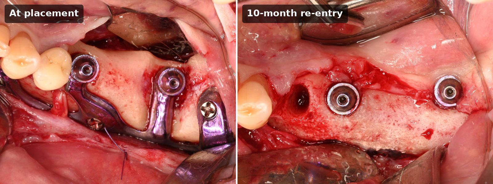
Key takeaways
- The main biological complication of modern subperiosteal implants is soft-tissue recession that exposes the framework.
- Gingiva lying directly on titanium has no vascular support from underneath; gingiva lying on bone does.
- The intraosseous concept combines guided bone preparation, bone regeneration over the framework, and two-stage, fully submerged healing.
- In an 88-year-old patient, a bone layer several millimeters thick covered the implant at 10 months, visible on CBCT and confirmed at surgical re-entry, with no soft-tissue recession.
Why soft tissue is the weak point of subperiosteal implants
With modern patient-specific subperiosteal implants, the framework itself is rarely the problem; the soft tissue around it is. In a multicenter study of 40 patients treated with additively manufactured subperiosteal implants in the maxilla, mucosal recession exposing the framework was found in 26 patients (65%), and both a thin soft-tissue biotype and mucositis were significantly associated with it [1]. Reported rates vary widely with the site, the implant design and the surgical protocol, which is precisely why the way the framework is covered deserves attention.
Two histological situations can be observed above the arm of a subperiosteal implant:
- No bone coverage. The gingiva lies directly on the titanium surface of the implant arm. Titanium is avascular, so the soft tissue receives no blood supply from underneath. A thin mucosa resting on a rigid, avascular surface is exposed to recession.
- Bone coverage. A layer of bone covers the implant arm. The gingiva rests on vascularized tissue that supports it from below. This is the biological rationale for a low risk of recession.
The intraosseous subperiosteal implant concept aims to move every case from the first situation to the second.
The intraosseous subperiosteal implant concept
The principle is easy to state. Instead of leaving the framework under the periosteum and the gingiva, the implant is planned and placed so that bone forms over it. The implant is subperiosteal by design but becomes intraosseous through healing. In practice, this relies on three elements:
- Guided bone preparation: a guide designed together with the implant is used to prepare the bone; in the case below, bony slots to receive the implant pillars.
- Bone regeneration over the framework: the implant is covered with a bone graft and a membrane, following the principles of guided bone regeneration.
- Two-stage, submerged healing: the implant is completely buried under the flap, with no healing caps, and uncovered only at the second surgical stage.
Biological rationale
The idea has experimental roots. In 1995, Hjørting-Hansen and colleagues fixed cast titanium frameworks on rabbit tibiae and covered half of them with an ePTFE membrane: regenerated bone partly covered the implants on the membrane side, whereas the control implants were mainly embedded in fibrous tissue [2]. In a follow-up study, the same group packed bovine bone substitute (Bio-Oss®) over the frameworks before covering them with a membrane. After 12 weeks, all 18 subperiosteal implants were completely osseointegrated, with most graft particles incorporated in newly formed woven bone [3].
Bone formation over a titanium device placed on the bone surface has also been reported in humans. Cohen et al. designed an additively manufactured, surface-modified Ti-6Al-4V subperiosteal device intended to generate bone; postoperative images at 3 and 8 months showed new bone formation and osseointegration of the device [4]. More recently, a preclinical study used 3D-printed polycaprolactone scaffolds specifically to obtain bone coverage over titanium subperiosteal implants [5], a sign that this goal is now being actively pursued.
Biomechanical rationale
Engaging bone around the framework may also matter mechanically. In a 2026 finite element study, Tang et al. modeled a hybrid design combining intraosseous fixtures with a subperiosteal titanium mesh in a severely atrophic mandible and compared it with configurations based on ultra-short implants. Under simulated masticatory loads, the hybrid implant remained well within the elastic range of the material, and the authors concluded that it offers superior biomechanical stability in severe mandibular atrophy [6]. The device studied differs from the one presented here, and finite element results are models rather than clinical outcomes, but they support the rationale for combining intraosseous engagement with a subperiosteal framework.
Clinical case: an 88-year-old patient with an atrophic posterior mandible
Initial situation
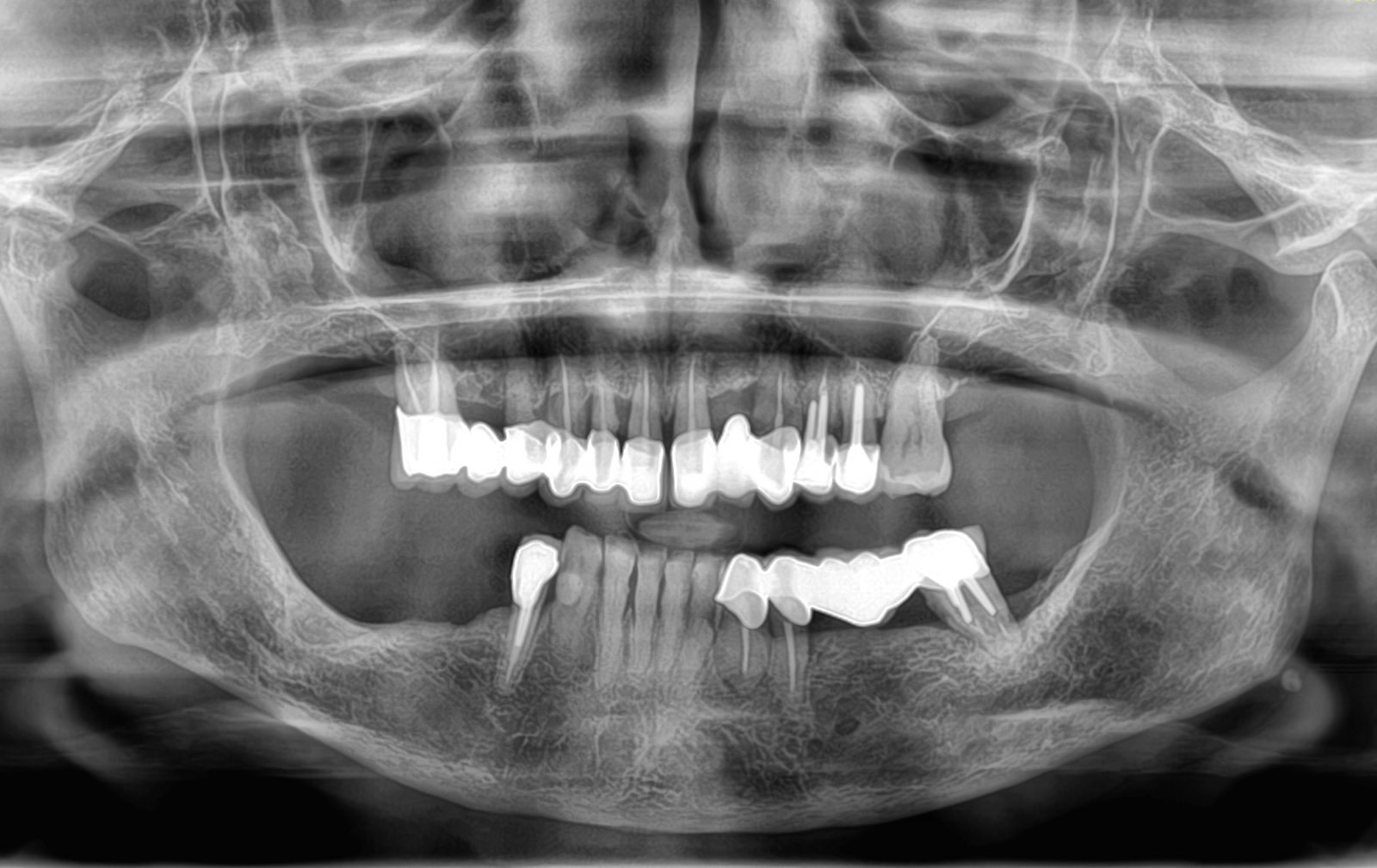
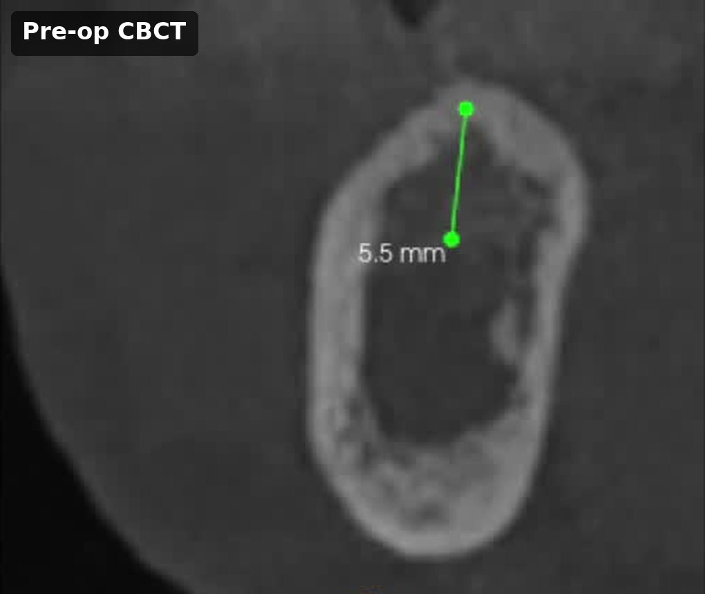
An 88-year-old woman presented with an edentulous, severely resorbed right posterior mandible. The preoperative CBCT showed only a few millimeters of bone above the inferior alveolar canal (5.5 mm on the cross-section shown below). At this age, osteogenic potential is reduced, which makes this case a demanding test for a concept that relies on bone regeneration.
Digital planning
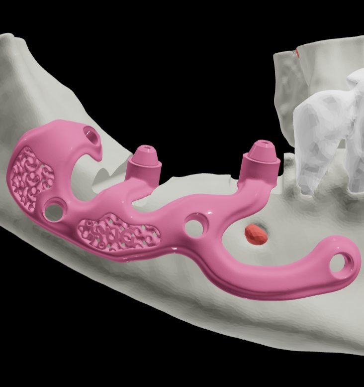
The implant was designed from the CBCT data as a patient-specific titanium framework. One of its arms was planned to pass below the mental foramen, and a bone-supported guide was designed to prepare the bone for the implant pillars.
First stage: placement under guided bone regeneration
The surgical sequence followed the same protocol we use in our other cases:
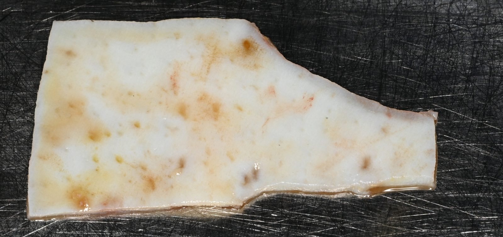
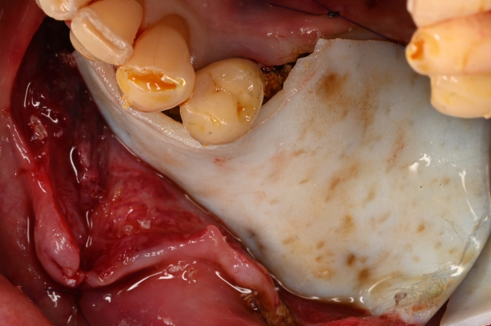
- A dermal collagen membrane was trimmed to the required shape.
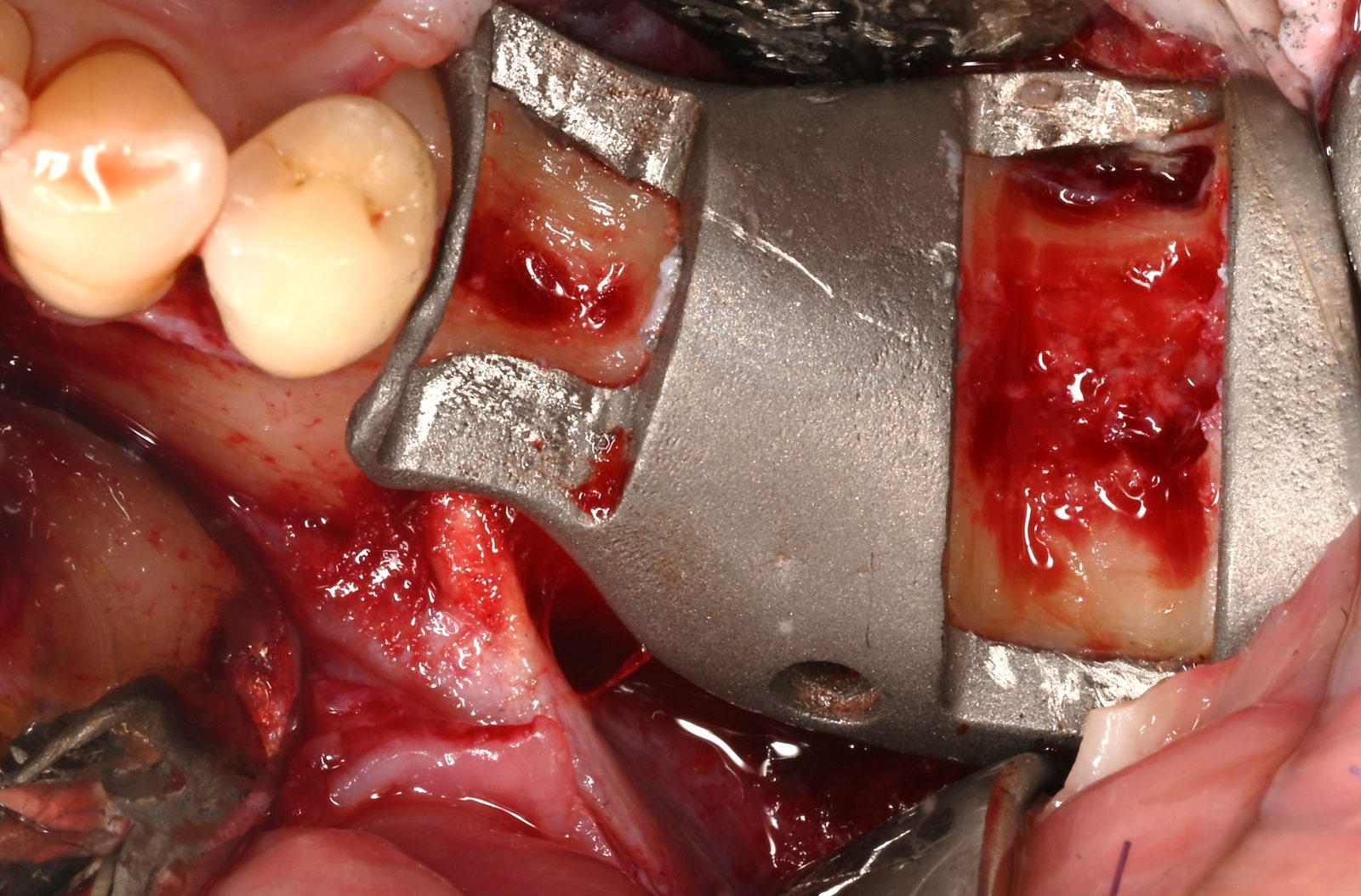
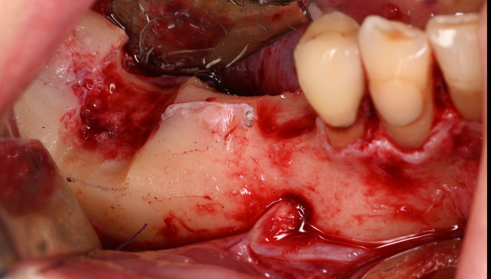
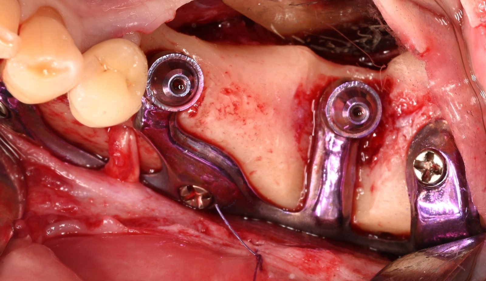
- After flap elevation, the guide was used to prepare bony slots for the implant pillars.
- The clinical situation matched the digital plan closely.
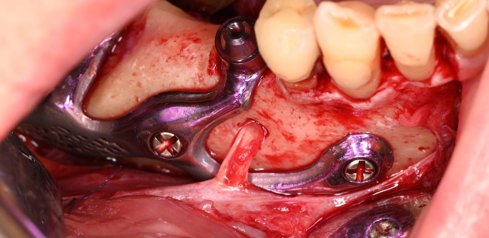
- The implant was seated, its arm passing below the mental foramen, and fixed with screws.
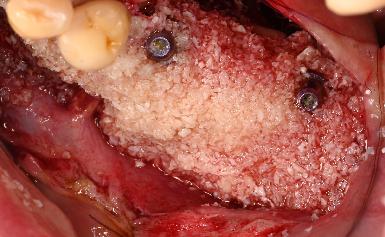
- The framework was covered with sticky bone, then with the membrane.
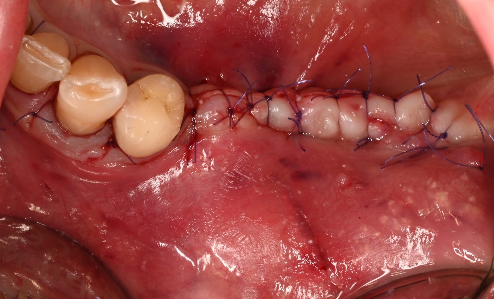
- The flap was closed over the entire construct. The implant was fully submerged, with no healing caps on the abutments: a two-stage approach.
Second stage at four months
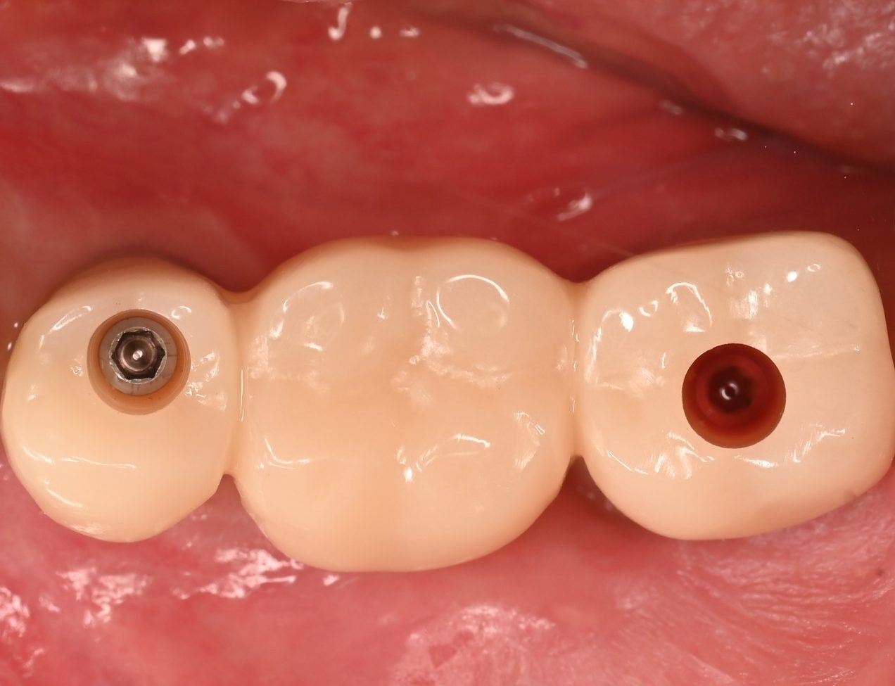
After four months, the mucosa had healed over the implant. The mesial abutment had become slightly exposed during healing, without any consequence. At the second surgical stage, the abutments were uncovered and fitted with healing caps; the screw-retained provisional bridge was placed two weeks later.
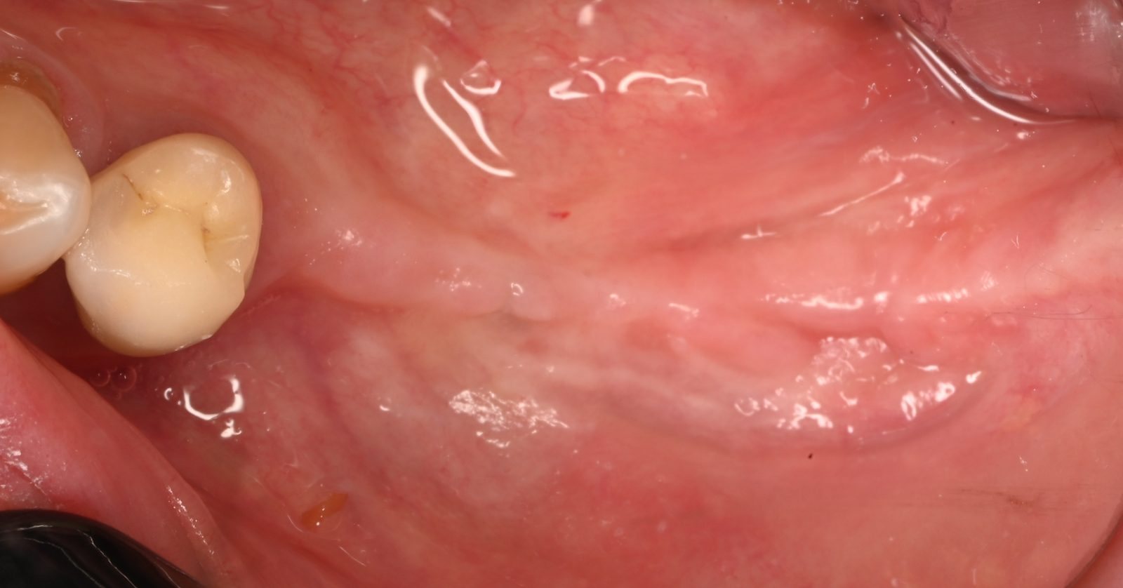
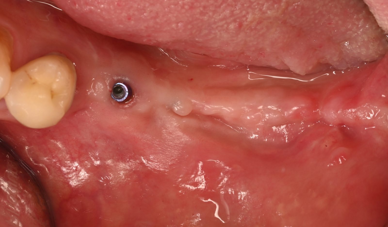
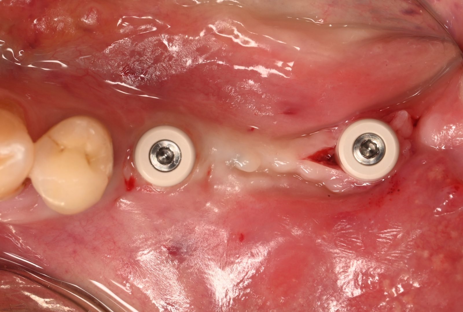
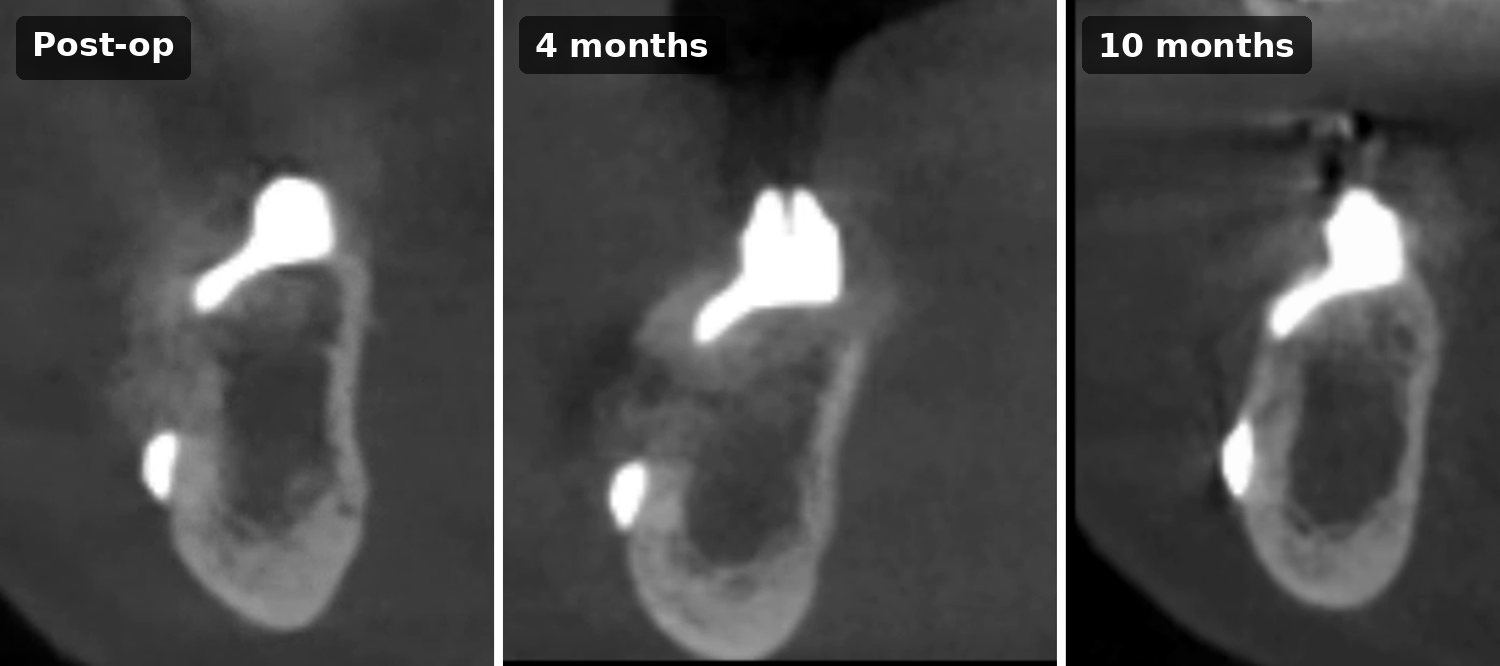
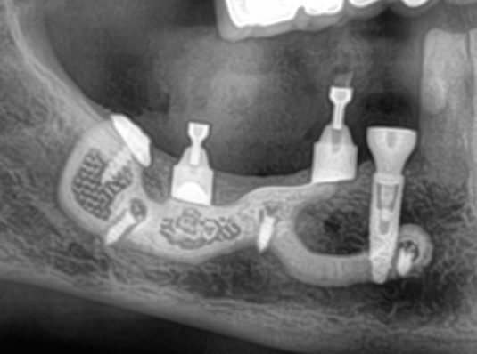
The CBCT comparison is the key finding at this stage. On the immediate postoperative scan, the graft over the framework has a heterogeneous appearance; at four months, it has densified, and bone height has increased compared with the initial situation. Radiographically, the framework is no longer lying on the bone but embedded in it: an intraosseous subperiosteal implant.
An unplanned re-entry at 10 months
Ten months after implant placement, the adjacent premolar (tooth 44, FDI) suffered a root fracture. Replacing it with a conventional implant required a surgical re-entry next to the subperiosteal implant, which gave us the opportunity to look directly at the framework.
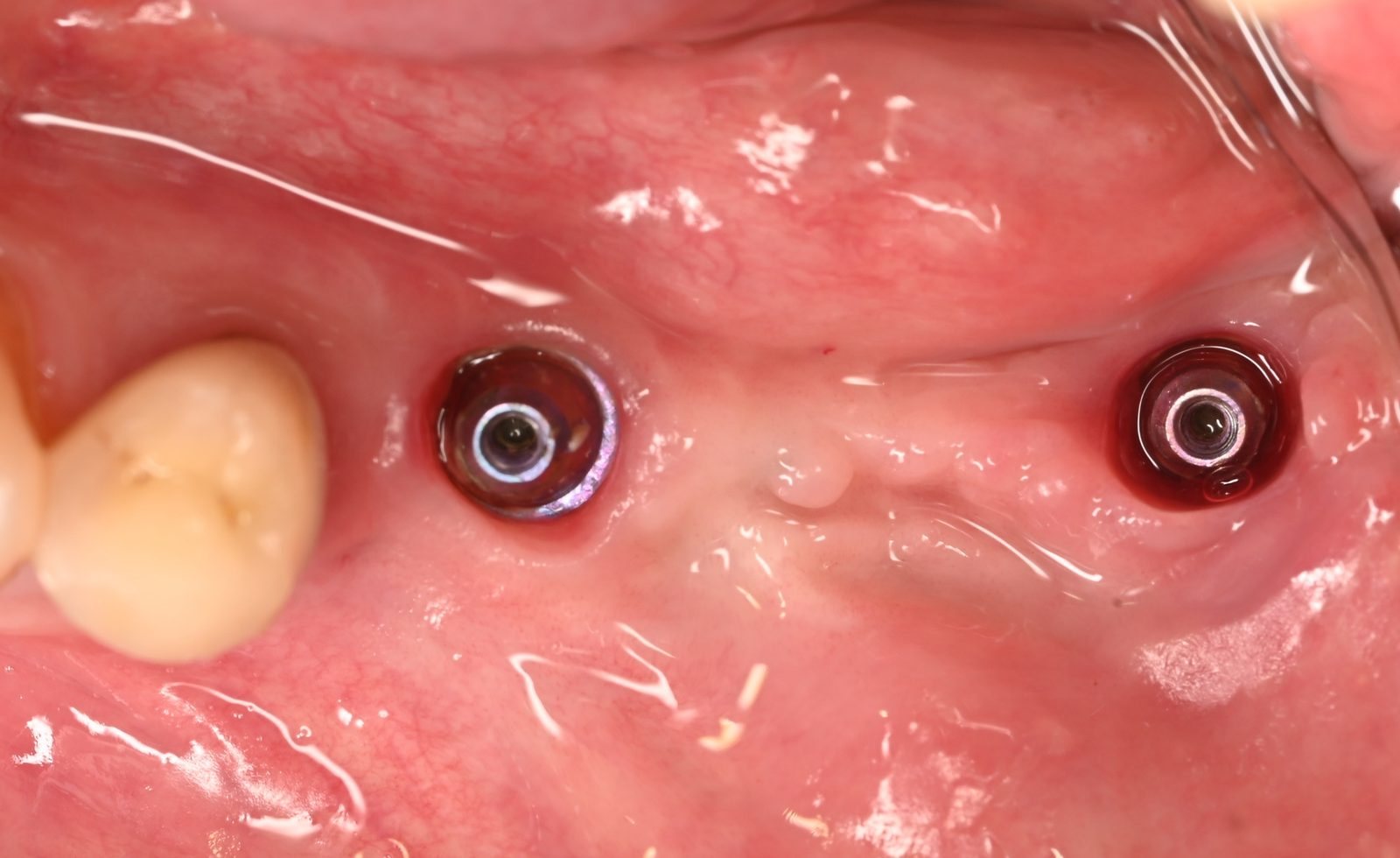
Before surgery, the soft tissues around the abutments showed no recession. At re-entry, the arms that were visible at placement were covered by a layer of bone several millimeters thick. The same bone layer was visible on the 10-month CBCT.
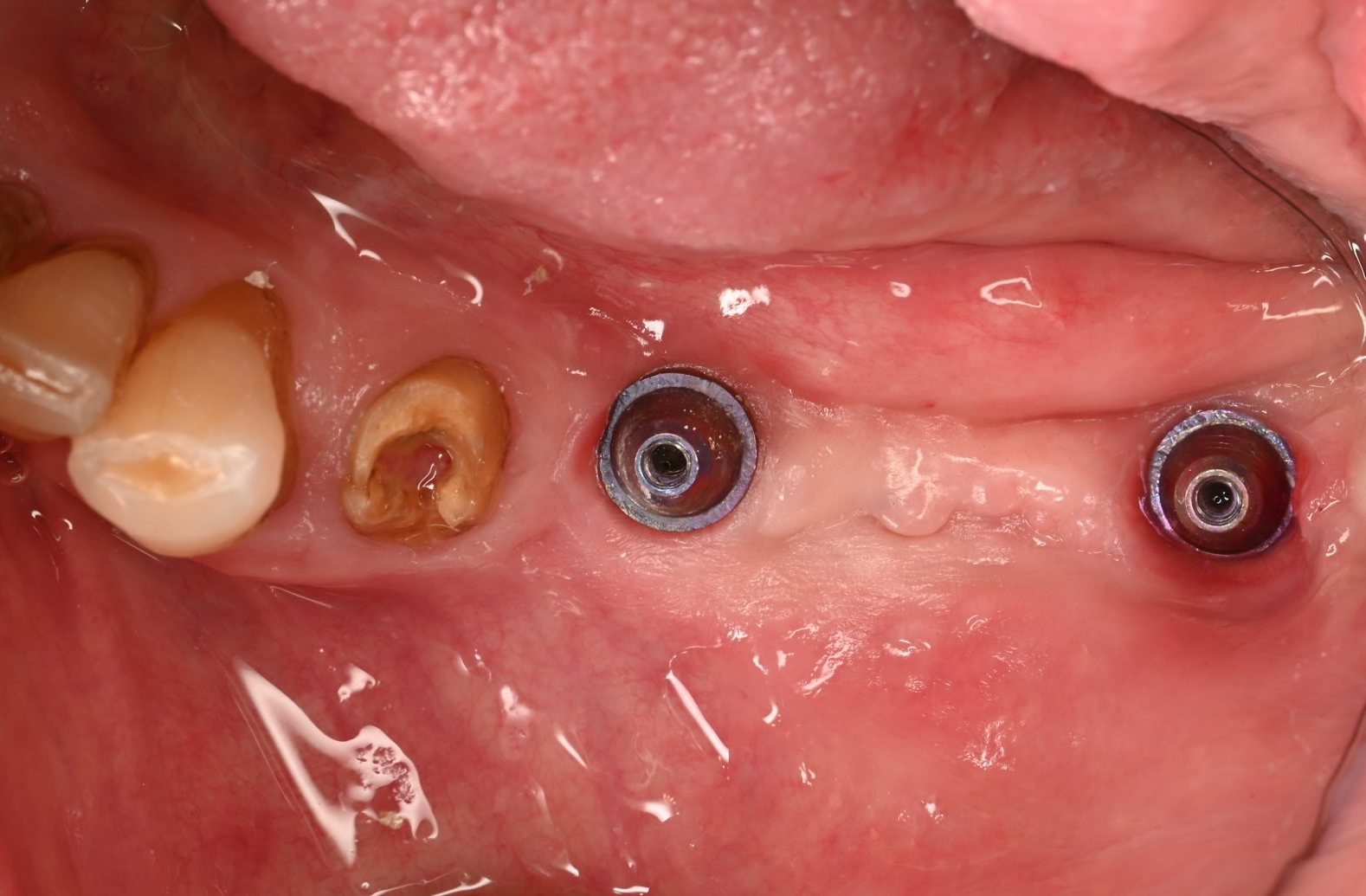
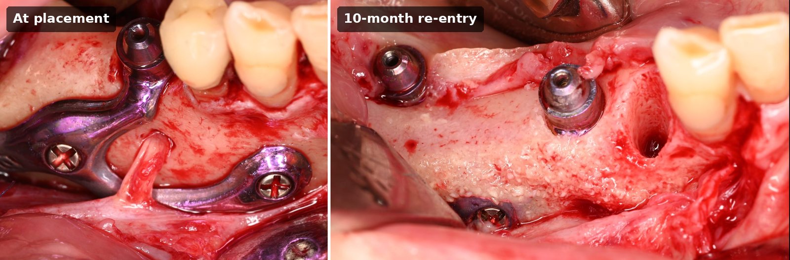
For the first time in our clinical experience, a substantial bone layer, several millimeters thick, was visible over the implant both on the follow-up CBCT and directly at surgical re-entry.
What this case shows, and what it does not
This case shows that bone coverage of a subperiosteal framework can be obtained even in an 88-year-old patient with a severely resorbed posterior mandible. The bone layer was not only a radiographic impression: it was observed directly at surgery. At 10 months, there was no soft-tissue recession around the abutments.
Its limits must be stated just as clearly:
- It is a single case, with a 10-month follow-up.
- There is no histology: we cannot state whether the regenerated bone is in direct contact with the titanium surface.
- CBCT around titanium is subject to metal artifacts, so radiographic bone over metal must be interpreted with caution. This is what makes the direct surgical observation valuable.
- Whether the bone layer remains stable over the long term, under function, still has to be documented in larger series with longer follow-up.
Key protocol points
- Design the implant and the bone preparation guide together, from the CBCT data.
- Prepare bony slots with the guide to receive the implant pillars.
- Fix the framework with screws.
- Cover the framework with a bone graft (here, sticky bone) and a collagen membrane.
- Close the flap over the entire construct: no healing caps, fully submerged healing.
- Uncover at four months: healing caps first, provisional bridge two weeks later.
A new perspective on subperiosteal implants
A subperiosteal implant does not have to remain under the gingiva. By combining guided bone preparation, bone regeneration over the framework and two-stage submerged healing, it can become intraosseous, giving the soft tissue the vascularized support it lacks over bare titanium. This case shows that the result is achievable even when regenerative potential is limited. The next step is confirmation in larger series and over the long term.
These principles (implant design, cutting guide and tissue management) are integrated into the HUG® concept, developed in collaboration with Biotech Dental®.
Learn the protocol in our training courses
Our on-site courses cover the fundamentals (bibliography, anatomy, finite element studies), digital design and workflow, surgical protocols for the maxilla and the mandible including the cutting guide, and provisional and final prosthetics, with a hands-on session.
Our e-learning (7 hours of online course) is free for practitioners who have attended an in-person training.
Frequently asked questions
What is an intraosseous subperiosteal implant?
A patient-specific subperiosteal implant that is planned and placed so that bone forms over its framework. It is subperiosteal by design and becomes intraosseous through healing, thanks to guided bone preparation, bone regeneration over the framework and two-stage submerged healing.
Why does bone coverage of the implant matter?
When the gingiva lies directly on titanium, it has no blood supply from underneath and is exposed to recession. When bone covers the implant arm, the gingiva rests on vascularized tissue.
How is the two-stage approach carried out in this protocol?
At placement, the implant is fully submerged under the flap with no healing caps. At four months, the abutments are uncovered and fitted with healing caps, and the provisional bridge is placed two weeks later.
References
- Van den Borre C, De Neef B, Loomans NAJ, Rinaldi M, Nout E, Bouvry P, Naert I, Van Stralen KJ, Mommaerts MY. Soft tissue response and determination of underlying risk drivers for recession and mucositis after AMSJI implantation in the maxilla. Int J Oral Maxillofac Implants. 2024;39(2):302-309. doi:10.11607/jomi.10490
- Hjørting-Hansen E, Helbo M, Aaboe M, Gotfredsen K, Pinholt EM. Osseointegration of subperiosteal implant via guided tissue regeneration. A pilot study. Clin Oral Implants Res. 1995;6(3):149-154. doi:10.1034/j.1600-0501.1995.060303.x
- Aaboe M, Schou S, Hjørting-Hansen E, Helbo M, Vikjaer D. Osseointegration of subperiosteal implants using bovine bone substitute and various membranes. Clin Oral Implants Res. 2000;11(1):51-58. doi:10.1034/j.1600-0501.2000.011001051.x
- Cohen DJ, Cheng A, Kahn A, Aviram M, Whitehead AJ, Hyzy SL, Clohessy RM, Boyan BD, Schwartz Z. Novel osteogenic Ti-6Al-4V device for restoration of dental function in patients with large bone deficiencies: design, development and implementation. Sci Rep. 2016;6:20493. doi:10.1038/srep20493
- Goh R, Vaquette C, Carluccio D, Ivanovski S, Ghosh Y, Breik O, Batstone M. Bone regeneration over subperiosteal implants using 3D-printed polycaprolactone scaffolds. J Craniomaxillofac Surg. 2026. doi:10.1016/j.jcms.2026.104590
- Tang Y, Nie H, Liu Z, Zhou W. Innovative design of a novel combined intraosseous-subperiosteal hybrid implant for severe atrophic edentulous dentition: a finite element analysis. BMC Oral Health. 2026. doi:10.1186/s12903-026-09126-4